E-ISSN:2349-3267
P-ISSN:2349-5499
RNI:MPENG/2017/74037

Research Article

Oxygen Therapy

Pediatric Review - International Journal of Pediatric Research

2025 Volume 12 Number 3 Jul-Sep
Publisherwww.medresearch.in

Retinopathy of Prematurity and Its Association with Oxygen Therapy in Preterm Low Birth Weight Babies

Mandal AA1*, Nahar K2, Hosneara3, Mazid AHMR4, Yesmin N5
DOI:https://doi.org/10.17511/ijpr .2025.i03.02

1* Abul Azad Mandal, Senior Consultant (Pediatrics), General Hospital, Gaibandha, Bangladesh.

2 Khairun Nahar, Junior Consultant (Pediatrics), General Hospital, Gaibandha, Bangladesh.

3 Hosneara, Junior Consultant (Pediatrics), Aminbazar 20 Bed Hospital, Savar, Dhaka, Bangladesh.

4 Abu Haider Md Raziul Mazid, Medical Officer (Pediatrics Haemato-Oncology), General Hospital, Gaibandha, Bangladesh.

5 Nafisa Yesmin, Assistant Professor (Pediatrics Haemato-Oncology), Rangpur Medical College, Rangpur, Bangladesh.

Background: Retinopathy of prematurity (ROP) is a preventable cause of blindness in premature, low birth weight (LBW) infants, driven by abnormal retinal vascular development. Oxygen therapy, though life-saving, is a key modifiable risk factor when unmonitored, leading to retinal damage through disrupted angiogenesis. ROP follows a two-phase pathogenesis involving initial vessel growth suppression due to hyperoxia, followed by hypoxia-induced neovascularization. In countries like Bangladesh, rising preterm survival without adequate neonatal care infrastructure has increased ROP incidence.

Methods: This cross-sectional observational study was conducted in the NICU of Pediatrics Dept., General Hospital, Gaibandha in Bangladesh to assess the prevalence of ROP and its association with oxygen therapy among 50 preterm, low-birth-weight infants. Eligible neonates (<37 weeks, <2500g, oxygen-exposed, and screened for ROP) were enrolled over a defined study period of January to June 2023. Data were collected using structured forms covering clinical, demographic, and treatment details. ROP screening followed standard protocols using indirect ophthalmoscopy. Oxygen therapy mode and duration were recorded. Statistical analysis was performed using SPSS v26.0, applying descriptive statistics and chi-square/Fisher’s exact tests, with significance set at p < 0.05.

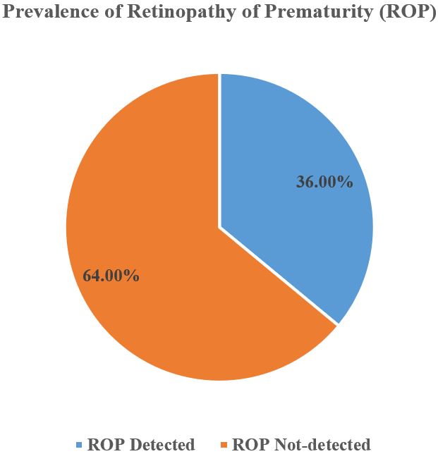
Results: Among 50 preterm low birth weight infants, 36% developed retinopathy of prematurity (ROP). Most were male (56%), born at 28–31 weeks (48%), and weighed 1000–1499 g (64%). CPAP was the most common oxygen modality (48%), with a mean oxygen duration of 5.8±2.3 days. ROP primarily involved Zone II (61.1%) and Stages 1–2. Bilateral ROP occurred in 61.1%, and 50% of cases required treatment. A significant association was found between oxygen therapy and ROP (p=0.04), especially with prolonged exposure (≥5 days; p=0.02) and mechanical ventilation (p=0.03), highlighting oxygen therapy as a key risk factor for ROP development.

Conclusion: This study highlights a significant association between unregulated oxygen therapy and ROP in preterm, low-birth-weight infants. Improved oxygen monitoring, adherence to saturation targets, and early ROP screening are essential to prevent vision loss. Strengthening neonatal care protocols can reduce the burden of this preventable cause of childhood blindness.

Keywords: Retinopathy of Prematurity, Oxygen Therapy, Preterm Infants, ROP Screening and Premature Birth

Corresponding Author How to Cite this Article To Browse
Abul Azad Mandal, Senior Consultant (Pediatrics), , General Hospital, Gaibandha, , Bangladesh.
Email:
Mandal AA, Nahar K, Hosneara, Mazid AHMR, Yesmin N, Retinopathy of Prematurity and Its Association with Oxygen Therapy in Preterm Low Birth Weight Babies. Pediatric Rev Int J Pediatr Res. 2025;12(3):63-69.
Available From
https://pediatrics.medresearch.in/index.php/ijpr/article/view/805

Manuscript Received Review Round 1 Review Round 2 Review Round 3 Accepted
2025-06-01 2025-06-07 2025-06-17 2025-06-27 2025-07-06
Conflict of Interest Funding Ethical Approval Plagiarism X-checker Note
None declared No funding sources Yes 11.55

© 2025 by Mandal AA, Nahar K, Hosneara, Mazid AHMR, Yesmin N and Published by Siddharth Health Research and Social Welfare Society. This is an Open Access article licensed under a Creative Commons Attribution 4.0 International License https://creativecommons.org/licenses/by/4.0/ unported [CC BY 4.0].

Download PDFBack To ArticleIntroductionMethodology and MaterialsResultsDiscussionConclusion and
Recommendations
References

Introduction

Retinopathy of prematurity (ROP) is a potentially blinding eye disorder that primarily affects premature infants with low birth weight (LBW). First described in the 1940s as “retrolental fibroplasia,” ROP has since become a significant cause of childhood blindness worldwide, particularly in countries with improving neonatal survival rates but limited access to comprehensive neonatal care [1],[2]. The disease is characterized by abnormal development of retinal blood vessels, often triggered by a range of perinatal factors, including oxygen therapy, sepsis, respiratory distress syndrome, and fluctuating blood oxygen levels [3].ROP predominantly affects infants born before 32 weeks of gestation or weighing less than 1500 grams at birth [4].

The immature retinal vasculature in these infants is highly susceptible to external influences, especially oxygen supplementation, which can disturb normal angiogenesis. During normal gestation, retinal vascularization is completed near term; however, preterm birth interrupts this process, making the retina vulnerable to pathological neovascularization in response to external stimuli like high oxygen concentrations [5].

Hence, while oxygen therapy is life-saving for preterm infants, it also poses a serious risk of developing ROP when not adequately monitored and regulated. The pathogenesis of ROP is understood to occur in two distinct phases. In the first phase, hyperoxia, often due to excessive supplemental oxygen, suppresses vascular endothelial growth factor (VEGF) and insulin-like growth factor-1 (IGF-1), halting normal vessel development. In the second phase, once oxygen levels normalize or drop, the retina becomes hypoxic, leading to a compensatory overproduction of VEGF, which stimulates disorganized and leaky neovascularization [6].

These pathological vessels can bleed, form fibrous scar tissue, and eventually cause retinal detachment and vision loss if not identified and managed early. Oxygen therapy has been consistently identified as one of ROP development’s most significant modifiable risk factors. Multiple studies have demonstrated a strong association between unregulated oxygen administration and increased incidence and severity of ROP [7].

The landmark Cryotherapy for Retinopathy of Prematurity (CRYO-ROP) and Early Treatment for Retinopathy of Prematurity (ETROP) trials underscored importance of early detection and treatment but also highlighted role of careful oxygen titration in preventing disease progression [8],[9]. Recent efforts, such as SUPPORT (Surfactant, Positive Pressure, and Oxygenation Randomized Trial) study, have attempted to refine optimal oxygen saturation targets to balance risks of ROP against those of other morbidities such as bronchopulmonary dysplasia and death [10].In resource-limited settings such as Bangladesh, burden of ROP is increasingly becoming apparent due to improved neonatal care and increased survival of preterm infants, but often without parallel implementation of screening and prevention protocols [11]. Many neonatal intensive care units (NICUs) lack stringent oxygen monitoring systems, exposing preterm infants to uncontrolled hyperoxia. This situation is exacerbated by inadequate training among healthcare providers and absence of national guidelines on oxygen therapy or ROP screening [12]. As a result, preventable cases of blindness due to ROP continue to rise. The global pattern of ROP is now described as occurring in three epidemic waves. The first was seen in 1940s and 1950s in high-income countries and was related to using unmonitored oxygen. The second occurred in 1970s and 1980s with improved neonatal survival. The third epidemic is currently unfolding in low- and middle-income countries, driven by increased access to neonatal care without proportional development of monitoring and screening infrastructure [13]. Given complex interplay between prematurity, low birth weight, and oxygen therapy in pathogenesis of ROP, this study aims to investigate association between oxygen therapy and development of ROP in preterm, low-birth-weight infants.

Methodology and Materials

This study employed a cross-sectional observational design conducted in the Neonatal Intensive Care Unit (NICU) of Pediatrics Dept., General Hospital, Gaibandha in Bangladesh. It aimed to determine the prevalence of retinopathy of prematurity (ROP) and evaluate its association with oxygen therapy among preterm low birth weight (LBW) infants. The study was conducted over 6 months, from January to June 2023. During the study period, 50 neonates were enrolled and analyzed in this study.


Inclusion Criteria:

  • Preterm neonates (<37 weeks of gestational age)
  • Birth weight less than 2500 grams
  • Admitted within 24 hours of birth
  • Received oxygen therapy during NICU stay
  • Underwent ophthalmologic screening for ROP

Exclusion Criteria:

  • Neonates with significant congenital anomalies
  • Neonates who died before ROP screening
  • Incomplete medical records
  • Parental refusal of participation

Data Collection Tools and Techniques:

Data were collected using a structured data collection sheet, which included the following variables:

  • Sociodemographic details: sex, gestational age, birth weight, mode of delivery
  • Clinical interventions: type and duration of oxygen therapy, pulse oximeter monitoring, surfactant therapy, presence of sepsis, and need for blood transfusion.
  • ROP-related findings: presence or absence of ROP, zone and stage classification, presence of plus disease, laterality, and treatment received

Gestational age was assessed using obstetric records and confirmed by clinical assessment (New Ballard Score when necessary). Birth weight was recorded using a calibrated digital scale within the first hour. Oxygen therapy details were recorded from NICU charts, including mode (nasal cannula, CPAP, mechanical ventilation) and duration (in days). Monitoring practices, sepsis diagnosis, and other interventions were extracted from patient records.

ROP Screening and Diagnosis:

All enrolled infants underwent ROP screening as per the hospital’s standard protocol:

  • The first ophthalmologic examination was performed between 4 to 6 weeks or at 31 weeks postmenstrual age, whichever was later.
  • Indirect ophthalmoscopy was performed by a trained pediatric ophthalmologist using a dilated pupil examination with a 20D lens.
  • ROP was staged and zoned according to the International Classification of Retinopathy of Prematurity (ICROP).
  • Follow-up examinations were conducted weekly or biweekly, depending on the stage and zone of disease.

Statistical Analysis:

Data were entered into Microsoft Excel and analyzed using SPSS version 26.0. Descriptive statistics such as frequencies, percentages, means, and standard deviations were used to summarize demographic and clinical variables. The chi-square or Fisher’s exact test (as applicable) was used to assess associations between categorical variables. A p-value <0.05 was considered statistically significant.

Results

The present study evaluated 50 preterm low birth weight babies to assess the association between oxygen therapy and the development of retinopathy of prematurity (ROP). As shown in Table 1, the study population consisted of 56% males and 44% females, with most infants born between 28–31 weeks of gestation (48%) and weighing between 1000–1499 g (very low birth weight, 64%). Cesarean section was the predominant mode of delivery (66%). Regarding oxygen support (Table 2), CPAP was the most commonly used modality (48%), followed by mechanical ventilation (28%) and nasal cannula (24%), with a mean oxygen therapy duration of 5.8±2.3 days. Continuous pulse oximeter monitoring was used in only 36% of cases, and comorbid conditions such as sepsis (42%), surfactant therapy (30%), and blood transfusion (26%) were also documented. Figure 1 reveals that ROP was detected in 18 infants (36%), while the remaining 32 (64%) showed no signs of the disease. Among those with ROP (Table 3), Zone II involvement was the most common (61.1%), and the most frequent stages were Stage 1 (27.8%) and Stage 2 (38.9%). Plus, the disease was present in 33.3% of the ROP cases. Bilateral involvement occurred in 61.1% of affected infants, and 50% required treatment. Among those treated, 55.6% underwent laser therapy, and 44.4% received anti-VEGF injections. A significant association was found between oxygen therapy and the occurrence of ROP (p=0.04), as shown in Table 4, where 40.5% of infants who received oxygen developed ROP compared to 12.5% who did not.


Table 5 further indicates that more prolonged oxygen exposure (≥5 days) was significantly associated with ROP development (72.2% vs. 20.8%; p = 0.02). Mechanical ventilation was statistically associated with ROP (66.7% vs. 33%; p=0.03), while CPAP and nasal cannula did not demonstrate significant relationships (p=0.08 and 0.12, respectively).

Table 1: Sociodemographic and Perinatal Characteristics of Preterm Low Birth Weight Babies (n = 50)

VariableFrequency (n)Percentage (%)
Sex of Baby
Male2856.00
Female2244.00
Gestational Age
<28 weeks612.00
28–31 weeks2448.00
32–36 weeks2040.00
Birth Weight
<1000 g (ELBW)510.00
1000–1499 g (VLBW)3264.00
1500–2500 g (LBW)1326.00
Mode of Delivery
Normal Vaginal Delivery1734.00
Cesarean Section3366.00

pediatric-805-01.JPG
Figure 1: Prevalence of Retinopathy of Prema-turity (ROP) Among Study Population (n = 50)

Table 2: Clinical and Supportive Care Interventions among Study Participants (n = 50)

VariableFrequency (n)Percentage (%)
Type of Oxygen Support
Nasal Cannula1224.00
CPAP2448.00
Mechanical Ventilation1428.00
Duration of Oxygen Therapy (Mean±SD)5.8 ± 2.3 days
Pulse Oximeter Monitoring
Continuous1836.00
Intermittent2040.00
Not Used1224.00
Surfactant Therapy1530.00
Sepsis2142.00
Blood Transfusion1326.00

Table 3: Distribution of ROP Characteristics, Laterality, and Management in Affected Infants (n = 18)

VariableFrequency (n)Percentage (%)
Zone of ROP
Zone I316.70
Zone II1161.10
Zone III422.20
Stage of ROP
Stage 1527.80
Stage 2738.90
Stage 3422.20
Stage 4211.10
Stage 500.00
Plus Disease633.3
Laterality
Unilateral738.90
Bilateral1161.10
Treatment Required
Yes950.00
No950.00
Treatment Given
Laser Therapy555.60
Anti-VEGF444.40
Surgery00.00

Table 4: Association between Oxygen Therapy and Occurrence of ROP (n = 50)

Oxygen Therapy GivenROP PresentROP Absentp-value
n%n%
Yes (n = 42)1740.52559.50.04*
No (n = 8)112.5787.5

Table 5: Association of Oxygen Duration & Mo-de of Delivery with Presence of ROP (n = 50)

VariableROP PresentROP Absentp-value
n%n%
Oxygen Duration ≥5 days1372.20927.800.02*
Oxygen Duration <5 days520.801979.20
CPAP (n = 20)630.001470.000.08
Mechanical Ventilation866.70433.000.03*
Nasal Cannula330.00770.000.12

Discussion

Retinopathy of prematurity (ROP) remains a significant cause of preventable childhood blindness, particularly in developing countries where advanc-ements in neonatal care have improved survival of premature infants, but not always with adequate monitoring protocols for ROP prevention. In this study, ROP was detected in 36% of 50 preterm LBW infants, aligning with prior findings in similar settings. Study in India reported an ROP prevalence of 30% among infants with comparable gestational ages and birth weights [14]. In contrast, higher-income countries report lower incidences, typically due to more stringent oxygen monitoring protocols and early screening practices [15]. This underscores importance of context-specific interventions in low-resource settings to mitigate preventable visual impairment. Our results demonstrate statistically significant association between oxygen therapy & ROP development. Among infants who received oxygen therapy (n=42), 40.5% developed ROP, whereas only 12.5% of those who did not receive oxygen showed signs of ROP (p=0.04). This finding is consistent with established understanding that unregulated oxygen supplementation is major modi-fiable risk factor for ROP. High oxygen concent-rations can disrupt retinal vascularization, causing vaso-obliteration followed by abnormal neovascul-arizationa hallmark of ROP pathogenesis [16,17]. WHO & American Academy of Pediatrics recommend maintaining oxygen saturation between 90-95% to balance risks of hypoxia & hyperoxia [18].

Unfortunately, only 36% of infants in this study had continuous pulse oximeter monitoring, highlighting a significant gap in care that may contribute to overexposure. The duration of oxygen therapy was also significantly associated with ROP. Infants receiving oxygen for ≥5 days had a markedly higher incidence of ROP (72.2%) compared to those receiving it for <5 days (20.8%), with a p-value of 0.02. These findings are corroborated by studies such as Dutta et al. [19], who reported a similar trend, emphasizing need to restrict duration of oxygen administration to only as long as clinically indicated. Notably, mechanical ventilation, a more invasive form of oxygen support, was associated with a significantly higher risk of ROP (66.7%) than those not ventilated (p=0.03). In contrast, CPAP and nasal cannula use did not show statistically significant associations, which may reflect differences in oxygen concentration and exposure intensity delivered by these modalities. Birth weight and gestational age were also critical in ROP risk stratification. The majority of infants with ROP were within very low birth weight category (1000–1499 g) and born between 28–31 weeks of gestation. This corresponds with existing literature, where immaturity of retina in lower gestational age infants predisposes them to vascular anomalies characteristic of ROP [20,21]. However, ROP was not exclusive to extremely low birth weight (ELBW) infants, suggesting that even moderately preterm infants are at risk when exposed to unregulated oxygen therapy. This finding reinforces need for universal screening guidelines tailored to local realities. The staging and classification of ROP in this study revealed that most cases were in Stage 1 (27.8%) and Stage 2 (38.9%), consistent with natural progression of disease in early detection settings. However, more severe stages like Stage 3 (22.2%) and Stage 4 (11.1%) were also observed, raising concern about delayed screening or rapid progression in some cases. Most ROP cases involved Zone II (61.1%), followed by Zone III and Zone I. These patterns align with Early Treatment for ROP (ETROP) study findings, which identified Zone I and posterior Zone II involvement as high-risk features [9]. The presence of plus disease in 33.3% of affected infants further underscores severity in a subset of cases. Treatment interventions were necessary for 50% of ROP-diagnosed infants, with laser therapy being predominant method (55.6%), followed by anti-VEGF therapy (44.4%).


These treatment modalities reflect global trends in ROP management, where anti-VEGF is increasingly used, especially in posterior ROP or when laser therapy is not feasible due to media opacity or systemic instability [22]. The absence of cases requiring surgery in this cohort suggests timely detection and management may have prevented progression to retinal detachment stages (Stage 5). Additionally, clinical comorbidities such as sepsis (42%), blood transfusions (26%), and the need for surfactant therapy (30%) were prevalent among the cohort, reflecting the multifactorial risk profile of these neonates. Prior studies have shown that systemic inflammation, oxidative stress, and transfusions contribute to the pathogenesis of ROP [23,24]. These associations further stress the importance of comprehensive neonatal care, including infection control, judicious transfusion practices, and regulated respiratory support.

Conclusion and Recommendations

In conclusion, the findings of this study reaffirm the strong link between unregulated oxygen therapy, exceptionally prolonged and high-concentration delivery, and the development of ROP in preterm, low-birth-weight infants. The results advocate for improved monitoring practices, including widespread use of continuous pulse oximetry and adherence to oxygen saturation guidelines, to prevent this avoidable cause of childhood blindness. Moreover, early screening and timely intervention remain critical components in managing ROP. Given the observed prevalence and treatment burden, this study highlights the urgent need to integrate ROP screening and oxygen regulation protocols into standard neonatal care practices in tertiary hospitals across Bangladesh.

Permission from Institutional Research Board: Yes

Funding: No funding sources

Conflict of interest: None declared

References

1. Tl T. Extreme prematurity and fibroblastic overgrowth of persistent vascular sheath behind each crystalline lens. I. Preliminary report. Am J Ophthalmol. 1942;25:203-4 [Crossref][PubMed][Google Scholar]

2. Gilbert C. Retinopathy of prematurity: a global perspective of the epidemics, population of babies at risk and implications for control. Early human development. 2008 Feb 1;84(2):77-82. [Crossref][PubMed][Google Scholar]

3. Fierson WM, American Academy of Pediatrics Section on Ophthalmology, American Academy of Ophthalmology, American Association for Pediatric Ophthalmology and Strabismus, American Association of Certified Orthoptists, Chiang MF, Good W, Phelps D, Reynolds J, Robbins SL, Karr DJ. Screening examination of premature infants for retinopathy of prematurity. Pediatrics. 2018 Dec 1;142(6):e20183061. [Crossref][PubMed][Google Scholar]

4. Blencowe H, Lawn JE, Vazquez T, Fielder A, Gilbert C. Preterm-associated visual impairment and estimates of retinopathy of prematurity at regional and global levels for 2010. Pediatric research. 2013 Dec;74(1):35-49. [Crossref][PubMed][Google Scholar]

5. Hartnett ME. Pathophysiology and mechanisms of severe retinopathy of prematurity. Ophthalmology. 2015 Jan 1;122(1):200-10. [Crossref][PubMed][Google Scholar]

6. Smith LE. Pathogenesis of retinopathy of prematurity. InSeminars in neonatology 2003 Dec 1 (Vol. 8, No. 6, pp. 469-473). WB Saunders [Crossref][PubMed][Google Scholar]

7. Chow LC, Wright KW, Sola A, CSMC Oxygen Administration Study Group. Can changes in clinical practice decrease the incidence of severe retinopathy of prematurity in very low birth weight infants?. Pediatrics. 2003 Feb 1;111(2):339-45. [Crossref][PubMed][Google Scholar]

8. Cryotherapy for Retinopathy of Prematurity Cooperative Group. Multicenter trial of cryotherapy for retinopathy of prematurity: preliminary results. Pediatrics. 1988 May 1;81(5):697-706. [Crossref][PubMed][Google Scholar]

9. Hardy R, Good W, Dobson V, Palmer E, Tung B, Phelps D. Early Treatment for Retinopathy of Prematurity Cooperative GroupRevised indications for the treatment of retinopathy of prematurity. Results of the early treatment for retinopathy of prematurity randomized trial. Arch Ophthalmol. 2003;121(12):1684-94 [Crossref][PubMed][Google Scholar]


10. SUPPORT Study Group of the Eunice Kennedy Shriver NICHD Neonatal Research Network. Target ranges of oxygen saturation in extremely preterm infants. New England Journal of Medicine. 2010 May 27;362(21):1959-69. [Crossref][PubMed][Google Scholar]

11. Ahmed AN, Muslima H, Anwar KS, Khan NZ, Chowdhury MA, Saha SK, Darmstadt GL. Retinopathy of prematurity in Bangladeshi neonates. Journal of tropical pediatrics. 2008 Oct 1;54(5):333-9. [Crossref][PubMed][Google Scholar]

12. Darlow BA, Hutchinson JL, Henderson-Smart DJ, Donoghue DA, Simpson JM, Evans NJ, Australian and New Zealand Neonatal Network. Prenatal risk factors for severe retinopathy of prematurity among very preterm infants of the Australian and New Zealand Neonatal Network. Pediatrics. 2005 Apr 1;115(4):990-6. [Crossref][PubMed][Google Scholar]

13. Graham EQ. Retinopathy ofprematurity: an epidemic in the making. Chinese medical journal. 2010 Oct 20;123(20):2929-37. [Crossref][PubMed][Google Scholar]

14. Vinekar A, Gilbert C, Dogra M, Kurian M, Shainesh G, Shetty B, Bauer N. The KIDROP model of combining strategies for providing retinopathy of prematurity screening in underserved areas in India using wide-field imaging, tele-medicine, non-physician graders and smart phone reporting. Indian journal of ophthalmology. 2014 Jan 1;62(1):41-9. [Crossref][PubMed][Google Scholar]

15. Quinn GE, Chan RP. Retinopathy of prematurity. InAlbert and Jakobiec's Principles and Practice of Ophthalmology 2022 Apr 23 (pp. 6737-6764). Cham: Springer International Publishing [Crossref][PubMed][Google Scholar]

16. STOP-ROP Multicenter Study Group. Supplemental therapeutic oxygen for prethreshold retinopathy of prematurity (STOP-ROP), a randomized, controlled trial. I: primary outcomes. Pediatrics. 2000 Feb 1;105(2):295-310 [Crossref][PubMed][Google Scholar]

17. Tin W, Gupta S. Optimum oxygen therapy in preterm babies. Archives of Disease in Childhood-Fetal and Neonatal Edition. 2007 Mar 1;92(2):F143-7. [Crossref][PubMed][Google Scholar]

18. World Health Organization. WHO recommendations on interventions to improve preterm birth outcomes. InWHO recommendations on interventions to improve preterm birth outcomes 2015. . [Crossref][PubMed][Google Scholar]

19. Dutta S, Narang S, Narang A, Dogra M, Gupta A. Risk factors of threshold retinopathy of prematurity. Indian pediatrics. 2004 Jul 1;41(7):665-72. [Crossref][PubMed][Google Scholar]

20. Gilbert C, Fielder A, Gordillo L, Quinn G, Semiglia R, Visintin P, Zin A, International NO-ROP Group. Characteristics of infants with severe retinopathy of prematurity in countries with low, moderate, and high levels of development: implications for screening programs. Pediatrics. 2005 May 1;115(5):e518-25. [Crossref][PubMed][Google Scholar]

21. Holmström G, El Azazi M, Jacobson L, Lennerstrand G. A population based, prospective study of the development of ROP in prematurely born children in the Stockholm area of Sweden. British journal of ophthalmology. 1993 Jul 1;77(7):417-23. [Crossref][PubMed][Google Scholar]

22. HA MH. Efficacy of intravitreal bevacizumab for stage 3+ retinopathy of prematurity. N Engl J Med. 2011;364:603-15. [Crossref][PubMed][Google Scholar]

23. Lee J, Chin JH, Kim JI, Lee EH, Choi IC. Association between red blood cell transfusion and long-term mortality in patients with cancer of the esophagus after esophagectomy. Diseases of the Esophagus. 2018 Feb 1;31(2):dox123. [Crossref][PubMed][Google Scholar]

24. Rivera JC, Holm M, Austeng D, Morken TS, Zhou T, Beaudry-Richard A, Sierra EM, Dammann O, Chemtob S. Retinopathy of prematurity: inflammation, choroidal degeneration, and novel promising therapeutic strategies. Journal of neuroinflammation. 2017 Dec;14:1-4. [Crossref][PubMed][Google Scholar]

Disclaimer / Publisher's Note The statements, opinions and data contained in all publications are solely those of the individual author(s) and contributor(s) and not of Journals and/or the editor(s). Journals and/or the editor(s) disclaim responsibility for any injury to people or property resulting from any ideas, methods, instructions or products referred to in the content.